Branch Retinal Vein Occlusion
Have you suffered a branch retinal vein occlusion? At Retina Associates of Middle Georgia, our team has served Middle Georgia since 1992. We are here to help you with your retina needs.
What is a Branch Retinal Vein Occlusion?
A branch retinal vein occlusion (BRVO) is a blockage of one of the retina’s smaller veins. These veins deliver blood to the retina. When they suffer a blockage, it leads to blood being unable to remove itself from the retina. As a result, fluid and blood leak from the retina’s blocked blood vessels. A retinal vein occlusion may lead to vision loss, especially if left untreated.
How Does a Branch Retinal Vein Occlusion Occur?
You may develop a branch retinal vein occlusion if the veins of your eyes are too narrow. Branch retinal occlusions occur when a blood clot blocks access to the vein. You’ll experience compression of these veins if an artery loses flexibility, or it becomes hardened.
Your likelihood of developing a branch retinal vein occlusion increases if you have high blood pressure, diabetes, high cholesterol, or other conditions that affect your body’s ability to control blood flow.
Are There Signs or Symptoms of a Branch Retinal Vein Occlusion?
If you have a retinal vein occlusion, it may not be something you realize you have right away. Some symptoms you may experience include:
• Painless blurring of your vision that gets worse over a period of hours or days
• Loss of vision, which may worsen slowly
• Complete loss of vision almost immediately
As these symptoms range from subtle to more apparent, it’s essential to schedule an appointment with your eye doctor as soon as possible if you experience them. If you have a branch retinal vein occlusion, leaving it untreated can lead to permanent retina damage and vision loss. It can also result in other eye problems.
How Do You Diagnose a Branch Retinal Vein Occlusion?
You may develop a branch retinal vein occlusion if the veins of your eyes are too narrow. Branch retinal occlusions occur when a blood clot blocks access to the vein. You’ll experience compression of these veins if an artery loses flexibility, or it becomes hardened.
Your likelihood of developing a branch retinal vein occlusion increases if you have high blood pressure, diabetes, high cholesterol, or other conditions that affect your body’s ability to control blood flow.
Are There Signs or Symptoms of a Branch Retinal Vein Occlusion?
If you have a retinal vein occlusion, it may not be something you realize you have right away. Some symptoms you may experience include:
- Painless blurring of your vision that gets worse over a period of hours or days
- Loss of vision, which may worsen slowly
- Complete loss of vision almost immediately
As these symptoms range from subtle to more apparent, it’s essential to schedule an appointment with your eye doctor as soon as possible if you experience them. If you have a branch retinal vein occlusion, leaving it untreated can lead to permanent retina damage and vision loss. It can also result in other eye problems.
How Do You Diagnose a Branch Retinal Vein Occlusion?
If your eye doctor thinks you have a branch retinal vein occlusion, they will do a series of tests and imaging to diagnose it, including:
Ophthalmoscopy
During ophthalmoscopy, your ophthalmologist will use a tool called an ophthalmoscope. The ophthalmoscope allows your eye doctor to see any changes to the retina that a branch retinal vein occlusion may have caused.
Optical Coherence Tomography (OCT)
Optical coherence tomography takes high-definition images of your retina with a scanning ophthalmoscope. With these images, your ophthalmologist at Retina Associates of Middle Georgia can see if there’s any swelling by measuring how thick your retina is.
The OCT images allow your eye doctor an accurate way to document the condition’s progress as you receive treatment for it.
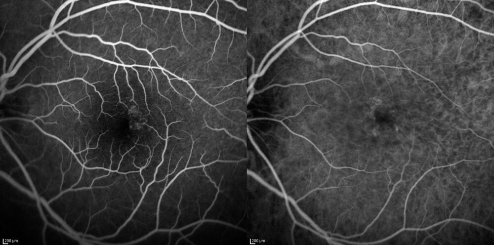
Fluorescein Angiography
Fluorescein angiography is a test procedure that uses dye injected into a vein in your arm. The dye eventually travels to the blood vessels in the retina.
With the dye, your ophthalmologist can see the blood vessels and take special photographs of them.
Can You Treat a Branch Retinal Vein Occlusion?
Although there is no way to unblock veins in the retina, your eye doctor can treat related problems due to the branch retinal vein occlusion. At Retina Associates of Middle Georgia, we treat this condition using anti-VEGF injections.
Anti-VEGF Injections
Anti-VEGF injections target VEGF, a growth factor that causes macular edema. Macular edema occurs when there is swelling in the macula due to blood and fluids leaking into it.
Anti-VEGF injections can help maintain or improve your vision after you’ve suffered a branch retinal vein occlusion. They help stop the growth of new abnormal blood vessels in the eye while also reducing the leaking of these vessels.
Focal Laser Treatment
If patients do not respond well to anti-VEGF injections, we may recommend using focal laser treatment as a secondary treatment. Focal laser treatment uses a laser to create tiny burns on areas around the macula. This may help improve your vision, but it is not guaranteed for all patients.
Learn more about branch retinal vein occlusion and treating it by scheduling an appointment at Retina Associates of Middle Georgia in Macon, GA.