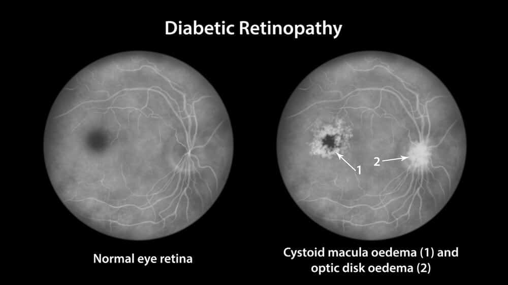
Cystoid Macular Edema
Have you learned that you have cystoid macular edema? At Retina Associates of Middle Georgia, our team of compassionate eye doctors is uniquely equipped to handle our patient’s retina treatment and care.
What is Cystoid Macular Edema?
Having cystoid macular edema means that the macula is swollen and filled with fluid. Anytime a part of the body becomes swollen, it’s known as edema.
If the macula fills with fluid because it’s swollen, it creates cyst-like patterns leading to cystoid macular edema. If you have cystoid macular edema, it affects your visual acuity, meaning your vision may no longer be as sharp as it once was.
You may suffer from blurry or wavy vision, especially in the middle of your field of vision. You may also notice that colors look different.
What Can Cause Cystoid Macular Edema?
Many things can potentially cause cystoid macular edema if your eye doctor at Retina Associates of Middle Georgia diagnoses you with the eye condition. Known causes that can lead to cystoid macular edema include:
• Being diabetic
• Undergoing eye procedures like cataract surgery or having a detached retina repaired
• Suffering a retina vein occlusion or blockage to the veins in the retina
• A side effect of medication you’re taking
• Injuring or hurting your eye
• You have eye inflammation
• You have age-related macular degeneration
Although these things can increase your risk of developing cystoid macular edema, it is not a guarantee that you will. Each patient and their eyes are unique.
How Do Eye Doctors Diagnose Cystoid Macular Edema?
If your eye doctor thinks you may have cystoid macular edema, they will run tests to confirm it, which may include the following:
Optical Coherence Tomography (OCT)
Your eye doctor uses a non-invasive test to diagnose cystoid macular edema with OCT. Optical coherence tomography uses a special kind of light that creates a high-definition cross-section image of the tissues in your eye, including your retina.
OCT is often thought to be one of the best ways of diagnosing cystoid macular edema.
Dilated Retinal Exam
During a dilated retinal exam, your ophthalmologist will dilate your eyes, allowing them to see your retina and your macula. They will use a special lens to see these parts of the eye and identify any cysts.
Fluorescein Angiography
Fluorescein angiography is a form of diagnostic testing that uses a camera to see any leaking blood vessels due to cystoid macular edema.
These are the three most common ways of diagnosing cystoid macular edema. Your eye doctor may use one or all of these diagnostic tools.
How Do You Treat Cystoid Macular Edema?
If you have cystoid macular edema, it’s necessary to see your eye doctor regularly. Regular appointments will ensure they can evaluate the condition’s cause and treat it accordingly.
Knowing the cause of cystoid macular edema will make it possible for your eye doctor to choose the best treatment. At Retina Associates of Middle Georgia, we treat cystoid macular edema using intraocular anti-VEGF injections and eye drops.
Anti-VEGF injections target VEGF in the retina. VEGF is responsible for the cyst-like growths found with macular edema.
We use anti-inflammatory eye drops to reduce swelling to improve your vision and visual acuity. As the edema goes away, you’ll need to continue seeing your eye doctor to ensure cystoid macular edema does not return.
Learn more about cystoid macular edema and if you may need treatment for this eye condition by scheduling an appointment at Retina Associates of Middle Georgia in Warner Robins, GA, now.